Get help now

Radiology of Pulmonary Infection, Lowry

  • Pages 3
  • Words 593
  • Views 494
  • Download

    Cite

    Bill
    Verified writer
    Rating
    • rating star
    • rating star
    • rating star
    • rating star
    • rating star
    • 5/5
    Delivery result 5 hours
    Customers reviews 893
    Hire Writer
    +123 relevant experts are online

    Academic anxiety?

    Get original paper in 3 hours and nail the task

    Get help now

    124 experts online

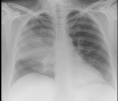
    Pneumonia CXR
    Pneumonia CXR
    Lung Cancer CXR
    Lung Cancer CXR
    Atelectasis CXR
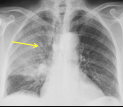
    RML.
    Right heart border is being blurred/lost
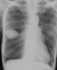
    Atelectasis CXR
    Loculated Pleural Fluid CXR
    This particular one is in the minor fissure
    Loculated Pleural Fluid CXR
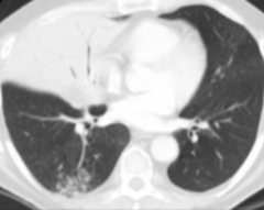
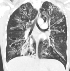
    Pneumonia CT
    Pneumonia CT
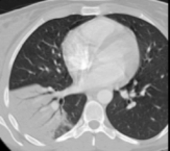
    Atelectasis CT
    Atelectasis CT
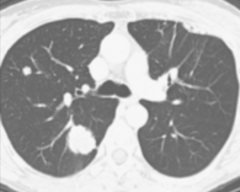
    Lung Cancer CT
    Lung Cancer CT
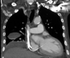
    Pleural Fluid CT
    Pleural Fluid CT
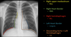
    The loss of a normal interface or border due to the pathological opacification of a region.
    Silhouette Sign
    Normal CXR Interfaces
    Normal CXR Interfaces
    Air Bronchograms
    -Accentuation of patent airways within an opacity
    -Surrounding airspace if fluid filled
    -More suggestive of pneumonia than atelectasis
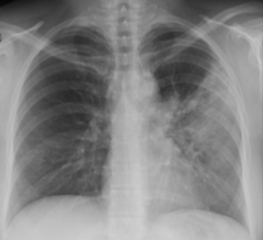
    Air Bronchograms
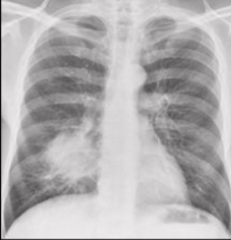
    Lobar Pneumonia CXR
    -Peripheral opacity that rapidly evolves into a confluent homogenous consolidation
    -Nonsegmental
    -Effects entire lobe
    -Commonly caused by streptococcus pneumoniae and klebsiella
    -usually heals without sequela
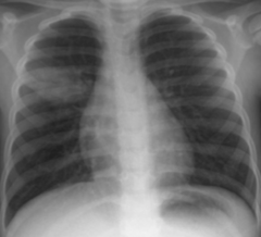
    Lobar Pneumonia CXR
    -Form of lobar pneumonia
    -Klebsiella classically, S. Pneumo more common
    -“Bulging fissure”
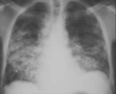
    “Round Pneumonia”
    Bronchopneumonia CXR
    -Infection of the airway mucosa that extends into alveoli
    -Patchy nodular opacities
    -S. Aureus, or G- organisms
    -Scarring after healing
    Bronchopneumonia CXR
    -Anaerobic bacteria
    -Findings; bilateral medial lower low basal segment, right more common than left.
    -Can become necrotic, capitate, and form an abscess.
    -Any patient that cannot protect their airway is at risk.
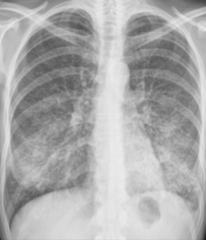
    Aspiration Bronchopneumonia
    Interstitial Pneumonia
    -Viruses, M. pneumoniae, PCP
    -Inflammation of interstitium
    -Bilateral symmetric linear reticular opacities
    -CT; Ground glass, whatever the &(%$ that is.
    Interstitial Pneumonia
    -Organizing pneumonia
    -Cancer
    -Timeline differentiates them.
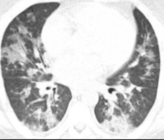
    Diseases that mimic pneumonia
    Organizing pneumonia
    -Disease with histo description of peripheral airspaces filling with mononuclear cells, foamy macrophages, and organizing fibrosis.
    -Many known causes
    -Findings; variable appearances with migratory multifocal peripheral opacities.
    -Clinical; patient with protected nonproductive cough and low grade fever with restrictive pattern on PFT. Does not respond to antibiotics, does respond to steroids.
    -Good prognosis.
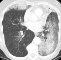
    Organizing pneumonia
    Cancer
    -Persistent opacity despite treatment
    Cancer
    Infectious nodules
    -Get smaller post treatment
    -Halo border
    Acutely present or subside on serial imaging
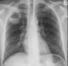
    Infectious nodules
    Lung Abscess
    -Thick walled cavity
    -Due to mixed anaerobic infection (S Aureus, pseudomonas)
    -Often related to aspiration, poor dental hygiene, LOC, esophageal dysmotlity, neurological disease
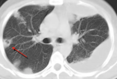
    Lung Abscess
    Septic Emboli
    -Hematogenous spread of infection
    -Multiple peripheral basilar nodules, which may cavitate.
    -Some may show a feeding vessel, and an infarct
    -Related to IVDU, and bacterial tricuspid valve endocarditis
    -Staph Aureus and epidermis.
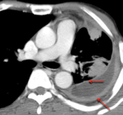
    Septic Emboli
    Empyema
    -Purulent material in the pleural space
    -Often related to evolution of a parapneumonic effusion, or an underlying lung infection that erupts into the pleural space (abscess or septic emboli).
    -Often located
    -Split Pleural sign
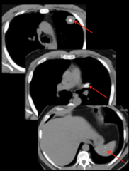
    Empyema
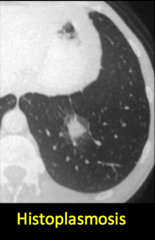
    Granulomatous disease in the lung
    -Granuloma; benign calcified nodules in the lung representing immune response to certain pathological insults.
    -Caused by infectious and non-infectious causes
    -Common infectious causes; Histo and TB.
    -Often seen with calcified hilar/mediastinal lymph nodes and hepatic/splenic granulomata.

    Granulomatous disease in the lung
    Progression of TB Infection
    Progression of TB Infection
    -Clinical infection following first exposure.
    -Usually asymptomatic in children, only detected via PPD.
    -Symptomatic in adults.
    -FTT, night sweats, weight loss, hemoptysis.
    -Often no imaging signs.
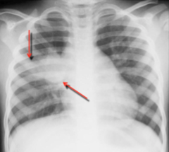
    Primary TB
    Findings in primary TB
    -Airspace consolidation, right more often than left.
    -Mediastinal and ipsilateral hilar lymphadenopathy in children and immunocompromised. Atelectasis may occur from compression of central airways.
    -Plural effusion, usually small, isolated, and unilateral.
    -Findings clear slowly.
    Findings in primary TB
    -Ghon complex; Visualization of sight of initial infection and enlarged ipsilateral lymph node.
    -Ranke Complex; Calcified tuberculoma and ipsilateral hilar lymph node.
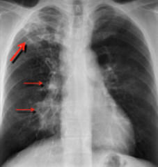
    Latent TB
    Primary Progressive TB
    -Consolidation process
    -Extensive consolidation and cavitation can develop.
    -Posterior upper lobe and superior segment of lower lobes is most common.
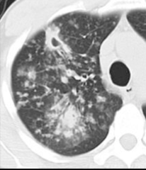
    Primary Progressive TB
    Post Primary TB
    -Reactivation TB
    -Classically in the apical posterior upper lobes and superior segments of lower lobes.
    -Rarely any pleural effusion or LAD.
    -May be associated with Tree in Bud opacities, which indicates the spread of the disease via the small airways. (Image)
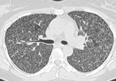
    Post Primary TB
    Disseminated Disease
    -Miliary TB
    -indicates hematogenous spread
    Disseminated Disease
    -CD4>200; typical post-primary findings
    -CD4<200; post primary resembles a primary infection; consolidation and LAD.
    Tuberculosis in the Immunocompromised
    -Consolidation
    -Endobronchial spread
    -Miliary Patterns
    -Centrilobular nodules (tree in bud)
    -Primary, progressive primary, post-primary.
    Signs of Active TB
    -Bronchiectasis
    -Linear scarring
    -Calcified nodules.
    -Stable for 6mos.
    Signs of inactive TB
    -M Avium Intracellulare Complex (MAC)
    -From natural water, soil, and animals.
    -Types; cavitary, bronchiectasis and nodules, centrilobular nodules.
    -Symptom; chronic cough.
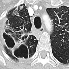
    Non-Tubercular mycobacterium
    Cavitary MAC
    -Resembles post primary TB
    -Older men in 60s with COPD or mildly immunocompromised.
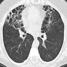
    Cavitary MAC
    Bronchiectasis and nodules MAC
    -Bronchiectasis with waxing/waning nodules.
    -Middle lobe and lingual predominant
    -Women in their 60s.
    -Lady Wndemere syndrome
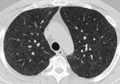
    Bronchiectasis and nodules MAC
    MAC with hypersensitivity pneumonitis
    -Centrilobular ground glass nodules
    -Owners of hot tubs
    -“Hot tub lung”
    MAC with hypersensitivity pneumonitis
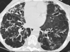
    Chronic Infection of the airways
    -Bronchitis; cough and fever, +/- consolidation
    -Bronchiectasis
    Chronic Infection of the airways
    Cystic Fibrosis
    -AR genetic disorder with decreased airway mucus clearance.
    -Upper lobe in central cystic/varicoid bronchiectasis
    -Pseudomonas, aspergillus, mycobacterial infection
    Cystic Fibrosis
    -Invasive; neutropenic patients.
    -Semi-invasive; mild immunocompromised patients. (Chronic necrotizing aspergillosis)
    -Mycetoma; normal immunity, history of apical cavity.
    -Findings; angio invasive (halos early, air crescent late), airway invasive (tree in bud and centrilobar nodules)
    Aspergillosis
    -Mild immunocompromised patients
    -Chronic necrotizing aspergillosis
    -Findings like TB; upper lobe consolidation and cavity.
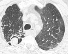
    Semi-invasive Aspergillosis
    Saprophytic Aspergillosis
    -Mycetoma.
    -Normal immunity
    -History of apical cavity (prior TB, bull, abscess)
    Fungus ball fills a preexisting cavity.
    Saprophytic Aspergillosis

    This essay was written by a fellow student. You may use it as a guide or sample for writing your own paper, but remember to cite it correctly. Don’t submit it as your own as it will be considered plagiarism.

    Need custom essay sample written special for your assignment?

    Choose skilled expert on your subject and get original paper with free plagiarism report

    Order custom paper Without paying upfront

    Radiology of Pulmonary Infection, Lowry. (2018, Oct 21). Retrieved from https://artscolumbia.org/radiology-of-pulmonary-infection-lowry-19129-59777/

    We use cookies to give you the best experience possible. By continuing we’ll assume you’re on board with our cookie policy

    Hi, my name is Amy 👋

    In case you can't find a relevant example, our professional writers are ready to help you write a unique paper. Just talk to our smart assistant Amy and she'll connect you with the best match.

    Get help with your paper